Cutting-edge flow, mass and imaging cytometry service and equipment
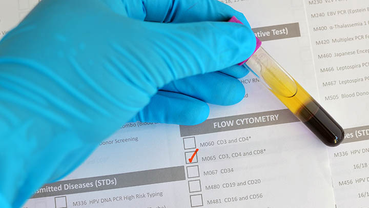
The Flow Cytometry Core Facility offers a suite of technologies to rapidly profile and sort large populations of cells and particles.
We offer researchers access to cutting-edge flow cytometry analysis and sorting equipment, high parameter mass cytometry and imaging cytometry.
In addition to The University of Manchester’s researchers, we also support external access.
How we work
Training and services
We combine training and service provision to researchers in order for them to perform high-quality multi-parameter flow, mass and imaging cytometry.
Training
We offer support and training on all aspects of flow cytometry from experimental design, basic operation and data analysis.
This training comes in the form of workshops, seminars, and one-to-one sessions.
We believe that having a good understanding of the fundamentals of a technique allows the users to get the most from the technology.
Services
Our staff are trained in the operation of highly specialised techniques such as cell sorting, mass cytometry and antibody conjugation.
Staff can run cell sorting, flow cytometry and mass cytometry projects for researchers that do not have the time or who are not fully trained.
The facility is managed full time by an experienced Senior Experimental Officer and is supported by three technical level staff.
Applications
Relevant experience and applications
We have extensive experience in a wide range of applications to support researchers across the University and beyond.
Applications include:
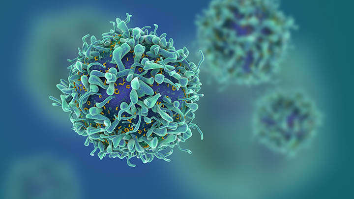
Immunophenotyping
High parameter immunophenotyping using fluorescence and mass cytometry (CyTOF) technologies.
Functional assays
Functional assays such as cell cycle, apoptosis, proliferation and cell signalling.
Small particle analysis
EVs and microparticles.
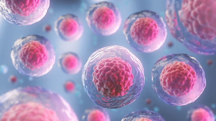
Cell sorting and cell line development
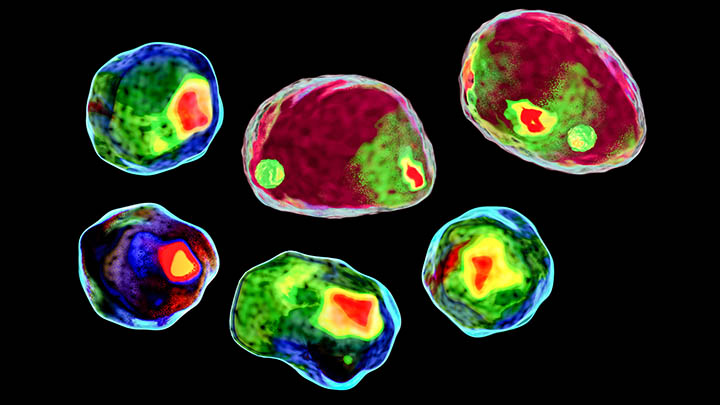
Imaging cytometry
Imaging cytometry with fluorescence (ImageStream) and mass-tagged antibodies (Hyperion).
Metabolomic studies (Seahorse)
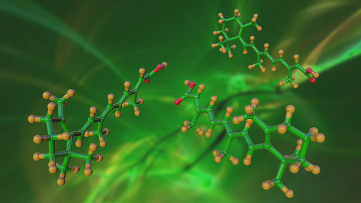
Antibody conjugation
Antibody conjugation for the development of reagents for CyTOF mass cytometry.
Technologies and equipment
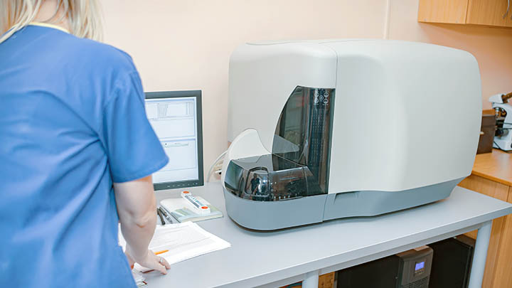
Our platforms
The Flow Cytometry Core Facility has a number of different platforms that enable a wide variety of applications to be undertaken.
Benchtop analysers
- BD FASC Verse (2-laser, 6 colour with plate reader)
- BD LSR-II (4 laser, 16 colour)
- BD Fortessa (4 laser, 17 colour)
- 2 BD Fortessa SORP (5-laser, 17 colour with plate readers)
- BD FACSymphony (5-laser, 28 colour with plate reader)
We have 6 analysers and 3 cell sorters in the flow cytometry facility capable of performing analysis and isolation of samples with up to 28 fluorescent markers. These machines allow the identification of a wide range of cell sub-types from a single sample thus increasing the magnitude of data obtainable from small and precious samples. Applications such as immunophenotyping, phosphoflow and functional assays are performed on these instruments. Soluble cytokine assays using capture beads can be run in 96 well plates on these platforms too.
Cell Sorting
- BD FACS Aria-Fusion (4 lasers, 4 way sorting, plates)
- BD Influx (5 laser, 6 way sorting, plates)
- Bigfoot (ThermoFisher) (7 laser, 6 way sorting, spectral, plates)
The cell sorters are able to isolate up to 6 separate populations of cells from a single sample and collect cells into tubes (bulk sorting) or as single cells into multiwell plates.
Cell sorting is performed by experienced facility staff which ensures the best quality of service and sort quality. Please contact us directly to arrange a sort or to discuss your requirements.
Spectral analysis and sorting
With the Bigfoot Cell Sorter we can perform both conventional and spectral analysis and cell sorting. While traditional filter-based cytometry cannot distinguish flurochromes with similar emission profiles, spectral cytometry enables the extension of panels using closely overlapping flurochromes due to its ability to separate emission signatures across the full spectrum. The Bigfoot has 60 individual channels and has the potential to expand investigative panels of markers to 40. In addition, the Bigfoot can use these large panels to identify and purify up to 6 populations simultaneously.
- Helios (Standard Biotools)
- CyTOF XT (Standard Biotools)
Mass cytometry, or CyTOF (cytometry by time of flight) is a platform that enables up to 50 individual markers or antibodies to be detected on a single cell suspension sample. Metal tagged antibodies are utilised in place of traditional fluorescent ones, and allied with mass spec detection, overcomes the intrinsic limitation to extended multi-antibody assays by reducing spill between channels to negligible levels. In conjunction with barcoding reagents, more than 500 samples can be run in a single tube using mass cytometry technology.
We offer an in-house custom conjugation service to label antibodies for use on the CyTOF and Hyperion (see below) platforms. We also offer ‘gateway’ panels of optimised antibodies in small aliquots to enable preliminary data to be collected at a cost effective price.
- Hyperion (Standard Biotools)
Using mass cytometry-based technology, the Hyperion imaging mass cytometer (IMC) is capable of imaging up to 50 antibody labels on a single tissue section. This significantly extends the ability to identify the cell types present within a single histology sample. IMC can be performed on both fresh/fixed and FFPE embedded tissue.
We offer an in-house custom conjugation service to label antibodies for use on the Hyperion and CyTOF (see above) platforms. We also offer ‘gateway’ panels of optimised antibodies in small aliquots to enable preliminary data to be collected at a cost effective price.
- Amnis ImageStreamX Mk-II (Luminex)
The ImageStreamX (ISX) is a combination of fluorescent microscopy and traditional flow cytometry. This platform supports your microscopy assays with high throughput imaging, over a thousand times faster than manual counting, by imaging up to 500 cells per second across 9 fluorescent channels. The ability to collect thousands of images enhances your quantitative assays. Additionally, it extends the assays available by flow cytometry with additional spatial analysis applications and can be used to identify where on or in your cells a drug delivery or bacteria may be, to visualise translocation events or cellular conjugation events.
The ISX has four objectives (20x, 40x and 60x) plus high sensitivity ‘High Gain’ and ‘deconvolution’ optics (EDF) for low signal amplification and spot counting respectively.
The ISX has a proven track record in characterising sub cellular and extracellular particles which are often on the scale of nanometres in size.
Publications and outputs
Impactful outputs
The Flow Cytometry Core Facility has been instrumental in research leading to top-class outputs.
Here are some of the recent highlights.
Med (N Y), 2021.
Utilising high parameter flow cytometry data and clustering analysis techniques, this paper details lymphocyte alterations in previously hospitalized COVID-19 patients up to six months following hospital discharge. It identifies three subgroups of convalescent patients based on distinct lymphocyte phenotypes, with one subgroup associated with poorer clinical outcome. The study indicates that alterations in B and T cell function following hospitalization with COVID-19 could affect longer-term immunity and contribute to some persistent symptoms observed in convalescent COVID-19 patients.
Bio Protocol, 2021.
We developed a method within the facility that allows for the analysis and selection of single fungal cells in high throughput by flow cytometry and fluorescence activated cell sorting (FACS), respectively. This method yields a throughput of thousands of cells per second. Selected cells can be subjected to downstream experiments to study single-cell behaviour.
Methods in Molecular Biology, 2021.
In collaboration with the Manchester Fungal Infection Group we developed a method using imaging flow cytometry to study the uptake of fungal spores on single human airway epithelial cells. In combination we used fluorescent dyes that could identify internalised and surface spores using the ImageStreamX. Using this technique researchers can study the molecular mechanisms of how individual cells respond to fungal infection and how anti-fungal treatments may be developed to utilise these mechanisms.
Journal of Experimental Medicine, 2021.
Utilizing a combination of high parameter flow cytometry analysis and cell sorting within the Facility, and services within the Bioimaging, Genomic Technologies, Bioinformatics and Biological Services core facilities at the University of Manchester this paper highlighted a novel pathway of myeloid cell development at a healthy barrier, defining a gingiva-specific stem cell network that supports generation of a proportion of the innate immune cells that police this barrier.
- DOI: 10.1084/jem.20200737
Science Immunology, 2020.
Utilizing a combination of high parameter flow cytometry analysis and cell sorting within the Facility, and services within the Bioimaging, Genomic Technologies, Bioinformatics and Biological Services core facilities at the University of Manchester this paper highlighted a novel pathway of myeloid cell development at a healthy barrier, defining a gingiva-specific stem cell network that supports generation of a proportion of the innate immune cells that police this barrier.
External access
Working with industry and other academic institutions
Although primarily focused on supporting research at The University of Manchester, we are also able to support work from other academic institutions and industry.
Academic institutions
We are happy to provide access to all of our systems to users from any academic institution.
We will provide these users with the same level of training and support as we would for our internal users.
Please contact us for further details.
Email: coreflowlab@manchester.ac.uk
Industry
We have worked for a number of multinational companies, both on an individual basis and as part of packages of work involving multiple facilities. Work may be on a collaborative basis or purely as a service provision.
All work is fully documented and contractual, and non-disclosure compliance is assured.
If you are interested in accessing the facility please contact our Business Development Manager for further details:
Dr Joanne Flannelly
Email: joanne.flannelly@manchester.ac.uk
Contact us
Find out more
Get in touch for further information or to inquire about using our facility.
Dr Gareth Howell, Senior Experimental Officer and Facility Manager
Email: coreflowlab@manchester.ac.uk
Tel: +44 (0)161 275 1742
Flow Cytometry Core Facility
Faculty of Biology, Medicine and Health
The University of Manchester
2.18d Core Technology Facility
Grafton Street
Manchester
M13 9NT
For news and updates from the facility, join us on our Slack channel.
Maps and travel
We are based in the Core Technology Facility (Building 86 on the University campus map).
Technology platforms
Technology platforms
We have a pioneering environment and facilities for research, innovation and technology development.
Technology platforms main page