Anatomy facilities
The University of Manchester has state-of-the-art anatomy facilities used by over 1,500 undergraduate students a year.
Our close collaboration with the onsite Manchester Surgical Skills and Simulation Centre brings together anatomy education for the next generation of healthcare professionals and scientists.
Our dedicated team of academics, technical staff and bequeathals administrators deliver teaching excellence through a diverse range of anatomical resources.
Dissection is an established teaching method that develops your 3D spatial understanding of the relationships between organs. It is the only instructional method that provides authentic haptic feedback, so it is crucial for future healthcare professionals to grasp.

You can witness anatomical variations firsthand and appreciate the uniqueness of each individual. This learning approach develops the vital constructs of professionalism, respect, morality, ethics and teamwork.
The dissecting room is a large, spacious facility where up to 40 body donors enable the teaching of topographical anatomy. Donors in the anatomy dissecting room are the embalmed bodies of public-spirited individuals who have left their remains to the University for medical and scientific teaching and research. Access to the dissecting room is regulated under the Human Tissue Act 2004.
Only students who take anatomy as a taught component of their course are granted access to the anatomy laboratory. You must first attend an introductory talk and sign a strict code of conduct before using the lab.
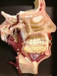
Students regularly use our extensive prosection collection to better understand human anatomy. Our collection of potted specimens demonstrates common pathologies that future clinicians and scientists may encounter.
Our vast collection of osteological material is used regularly by students to better understand surface landmarks through the study of bones combined with how bones appear on clinical images.
We have extensive archaeological skeletal material that is used to illustrate pathologies and kindle curiosity within our students.
Anatomy academics and technical staff work collaboratively with NHS colleagues to underpin our teaching with clinical relevance. We have a dedicated ultrasound room with 20 handheld scanners and two larger, but portable, ultrasound machines. Used in conjunction with other anatomical teaching methods, ultrasound enables our students to consolidate their knowledge and upskill in key techniques used regularly in the clinical setting.
Your understanding and interpretation of living anatomy will be strengthened by the inclusion of X-radiographs, CT and MRI scans alongside the use of wet specimens.
We strongly believe in catering for all learning styles and providing different methods of interacting with anatomy. Glasses-free 3D displays of the body, alongside Anatomage tables, offer different ways of learning. You'll also benefit from the University's subscriptions to Complete Anatomy and Acland’s video atlas of anatomy, which enables students to continue their anatomy studies away from campus.
Our digitised collection of histology slides, which can be viewed any time and from any place, will also enable you to learn more about macro and microscopic anatomy to further cement your understanding of this area of study.
