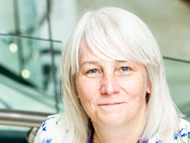
Using nanotechnology for targeted delivery
As Professor of Experimental Therapeutics, Kaye Williams' research involves exploiting nanotechnologies in different ways across her research programmes in the Division of Pharmacy and Optometry. Here, Kaye gives us an overview of how they are being used.

We exploit nanotechnologies in different ways across our research programmes, and have developed approaches that have enabled specific characteristics of nanotechnologies to be evaluated non-invasively, supporting the research of teams across nano-medicines and materials science.
It's a truly multidisciplinary, team science approach with collaboration and shared goals underpinning the research taken forward.

Professor Kaye Williams
Kaye Williams is Professor of Experimental Therapeutics at The University of Manchester and pre-clinical lead for oncology imaging within the Manchester Cancer Research Centre (MCRC).
Patient benefit
We've many new molecules that have potential as anti-cancer therapies, but they often don't possess all of the characteristics that are needed for success as a clinically used cancer drug. They may not accumulate sufficiently within the tumour to cause the desired effect, they may be excreted from the body too quickly, or they may cause unwanted toxicities.
One of the main aims of cancer-associated nanomedicine development is to generate therapies with improved drug targeting to cancer cells. Nano-carriers can provide a vehicle in which to encapsulate drugs and enable selective delivery to cancer cells.
Hopefully, by doing this, you increase the chance of your approach having an impact on the cancer cells while reducing the chances of it affecting other cells in the body and causing toxicity. That's certainly the approach we've been trying.
We're using nanomedicines to try and target the delivery of novel cancer therapeutics to specific types of cancer cells that are thought to be associated with poor treatment response. We're exploiting nano-carriers designed to bind to particular molecules that are expressed on the cell surface of cancer cells to selectively deliver novel therapies that prevent tumour growth.
We have been able to show that we can manipulate the interaction between hyaluronic acid and the receptor CD44 that's on cancer cells to deliver specific targeted molecules.
We have also been involved in research to develop additional approaches whereby the nano-carriers respond to specific cues” within the tumour microenvironment, and only release their drug cargo when they encounter the tumour-specific cue.
“For me, it's all about improved outcomes.”
Hypoxia and the tumour microenvironment
One key research area is hypoxia in tumours. This is a condition that naturally arises in all solid tumours. The problem, however, is that it causes resistance to therapies such as radiation treatment and can cause resistance to standard chemotherapy agents.
This is problematic, because it links with poor patient outcomes and with development of metastases and aggressive disease.
It can be challenging to deliver drugs to hypoxic regions, so what we want to be able to do is selectively deliver new therapies to hypoxic cells in as safe a way as possible for patients.
In the context of a nanomedicine approach, you can potentially exploit hypoxia as a cue for a targeted drug release, or use characteristics specific to hypoxic cells that enable selective delivery, which forms part of the research that we're trying to do.
In material science, we're generating specific materials to mimic the tumour micro-environment more rigorously than our standard cancer cell culture conditions, and provide an environment where the cancer cells start to behave more like they would do naturally within the body.
This helps us if we're screening new drugs, or trying to understand cancer evolution or processes such as metastasis, when cancer cells develop the ability to invade different tissues which are regulated via the interaction of cancer cells and their immediate environment.
Challenges
One challenge from this is being able to selectively deliver drugs to tumours, because even when we develop drugs that we think should target a biology that's specific to the tumour, you still have associated side effects. It's not enough that you're trying to target a tumour-specific characteristic; on its own, that won't prevent any potential toxicity.
Of course, classic standard chemotherapy agents that we're using routinely have considerable side effects associated with them, but they're very good at killing tumour cells.
Even routine chemotherapy agents could be delivered better. One important aspect from the nanotechnology side is to get a much more targeted delivery of drugs where you want, over the timeframes that you want.
There's huge potential in the application of nanotechnology to bridge these challenges, with work in many groups pushing towards the development of systems that can release therapies over time only under specific conditions to targeted sites. It's a very complex area, but significant advances are being made.
For me, it's all about improved outcomes, and not just improved outcome from the tumour response; it's reducing toxicity and having therapies going into patients that have a much improved safety profile while maintaining excellent anti-cancer effects.
“Manchester has expertise across a huge range of areas.”
Imaging
Part of my role within the Manchester Cancer Research Centre is to lead on preclinical imaging and, through this role, I work with a team of talented researchers who can develop novel means of non-invasively tracking labelled molecules or cells.
In cancer, and many other areas of research, we need to be able to visualise what's going on inside the body in real time and imaging allows us to do this. Two routinely used methods are positron emission tomography (PET) and magnetic resonance imaging (MRI). Our imaging team has supported nanotechnology research in both areas.
PET is one of the most sensitive imaging techniques. Many patients will have a PET scan, most commonly using the PET-tracer 18F- fluorodeoxyglucose - FDG. This is glucose labelled with a positron emitting radionuclide, which indicates where a cancer is in the body because of the increased uptake of glucose by cancer cells.
Similarly, you can label other types of molecule and see where they end up within the body. Dr Mike Fairclough has led our research linking non-invasive imaging approaches to nanotechnologies and materials science.
We worked with Professor Alberto Saiani to use PET-labelling of hydrogel molecules, developed as biomaterials for use in tissue engineering, to investigate how they behaved when administered in vivo.
We've also used different types of nanotechnologies, such as nano-rods, which are visible by MRI. Again, we've tagged those with PET tracers, so that enables you to develop brand new imaging agents for application in both MR and PET.
Cancer treatments are not only about using drugs. Increasingly, cells can be used as a potential therapy within patients.
Another application bridging nanotechnologies and imaging was to design a means of labelling cells to enable them to be tracked by PET. This would allow you to visualise where the cells end up when administered to a patient.
Here, a nano-carrier was loaded with a positron-emitting radionuclide and used to deliver the radionuclide to cells that we know are important in cancer biology and therapy response. This technology would allow us to then track those cell populations when we introduce them into a pre-clinical animal disease model, or eventually into patients.
Graphene and Manchester
Obviously, graphene has lots of potential applications in healthcare, but what we really need to know is where it might accumulate in a body when it's given.
You might want to test whether it's accumulated in a specific place but, equally, you may want to know how the body handles graphene. Does it accumulate in specific tissues that might actually cause a problem down the line, and how can we track that?
To do that, working with Professor Kostas Kostarelos' group, our imaging team have labelled different types of graphene with positron-emitting radionuclides that allow us to track it in vivo via PET imaging.
They were then able to track the bio-distribution of the different graphene molecules within the body and monitor how formulation affected tissue accumulation and excretion of the graphene in the urine.
There's obviously a lot of interest in Manchester about the applications of graphene in healthcare, and these types of studies showing how graphene distributes in the body and whether there would be any potential toxicity risk associated with this are very important.
Multidisciplinary research
I'm based in the Division of Pharmacy and Optometry, so I spend my time surrounded by colleagues who do everything from developing new drugs and nanotechnologies through to influencing policy and practice.
Manchester has expertise across a huge range of areas, and the research within my team is heavily reliant on collaborative networks that bring in expertise across many different areas.
Imaging is a great example here, as we are always aiming to translate our findings from bench to bedside. We want to be able to track how well the anti-cancer approaches we are developing are working as quickly as we can, not only from the perspective of our research questions, but also in terms of refining the work we do.
That's incredibly important research, and of course the types of imaging that I've focused on here are ones that are immediately translatable into patients.
If we're thinking about drug targeting, then we have a means of tracking that it has been successful through an imaging-based approach that has been developed in parallel, so when we go into our clinical situation, we're in a much more powerful position to be able to evaluate drug-response in patients quickly.
That's where the multidisciplinary work is coming from. I'm not a chemist so if I think we've got a good target in cancer, I liaise with my colleagues who would develop the potential drug to hit that target.
We then may need input on formulation, and targeting approaches afforded by nanotechnologies. I can evaluate these approaches, and we can look for developing biomarker and imaging profiles that would help us define response and select the right patients for the approach.
With the expertise in Manchester, we can cover all of these aspects in a coordinated approach. That's a really exciting environment to be in.
Learn more about nano-oncology research at the Manchester Cancer Research Centre.
